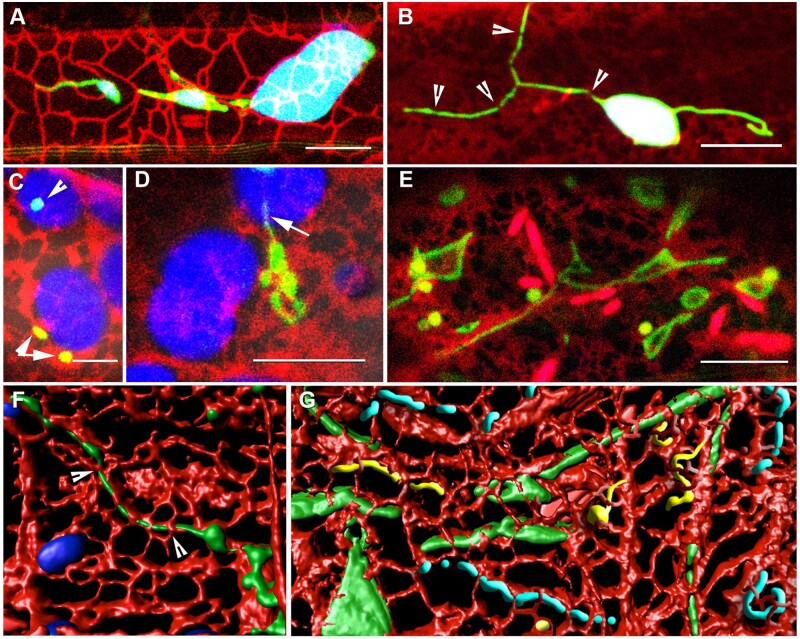
Figure 2.
The ER plays a major role in modulating the form and behavior of tubules. (A) Plastids surrounded by a loose ER cage (white outlined tubules against chlorophyll [blue] background) and stromules (green) aligned with red fluorescent protein-labeled ER (Supplemental Movie S4). (B) Dilations formed on a stromule due to constrictions (arrowheads) created by the ER mesh. The stromule extended on the other side remains free of constrictions and dilations. (C) Peroxisomes near chloroplasts (arrowheads) are actually embedded in the loose peri-plastidic ER mesh (red) suggesting that peroxisomal response to ROS generated by chloroplasts might be mediated by through the ER. (D) The ER–peroxule relationship illustrated by a peroxule (arrow) extended along an ER tubule (red) that is part of the ER cage around a chloroplast. (E) Expanded ER cisternae (red) and giant mitochondria (green) formed in epidermal cells in 7-d-old Arabidopsis seedlings after being kept in water under a coverslip for 45 min to create hypoxic conditions. (F) 3-D volume rendering of a confocal x, y, z image stack of an elongated peroxisome from the apm1 mutant and the surrounding ER (and blue rendered chloroplasts) suggests that tubular organelles are interspersed through the ER mesh. (G) A composite diagrammatic rendition suggests that tubular extensions from chloroplasts (green), peroxisomes (yellow), and mitochondria (blue) are all enmeshed in the ER (red). The dynamic behavior of the ER is thus responsible for the continuous shape changes of these organelles. Scale bars in µm: A, B, D, and E = 10; C = 5.

