Figure 2.
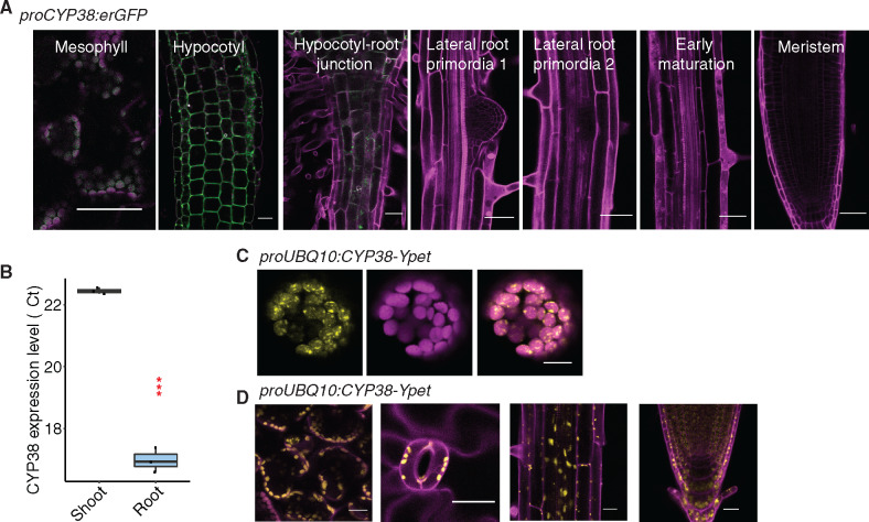
CYP38 is highly expressed in shoots and localized in plastids. A, Confocal images of proCYP38:erGFP transgenic line. Magenta indicates autofluorescence of chloroplasts in mesophyll cells together with PI staining in the hypocotyl and various regions of the root, while green indicates GFP fluorescence. Scale bars = 50 µm. B, Quantitative RT-PCR of CYP38 transcript level in WT shoot and root tissue. The numbers are presented as log2 transformed values (ΔΔCt). (***P < 0.001, pairwise t test, n =3). C, Confocal images of WT protoplast that was transiently transformed with proUBQ10:CYP38-Ypet. Magenta indicates chloroplast autofluorescence, while yellow indicates Ypet fluorescence. Scale bars = 10 µm. D, Confocal images of proUBQ10:CYP38-Ypet transgene fluorescence in various tissues including (left to right) mesophyll, stomata, mature root and root tip. Magenta indicates PI staining and autofluorescence from chloroplasts while yellow indicates Ypet fluorescence. Scale bars =20 µm.

