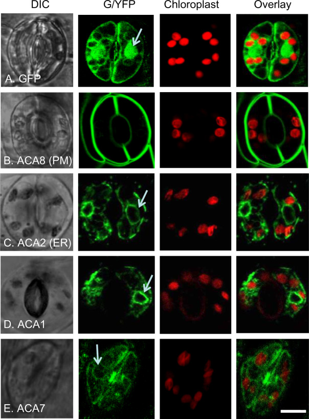
Figure 2.
Confocal fluorescence microscopy of guard cells showing FP-tagged ACA1 and 7 with subcellular localizations similar to ER-localized ACA2. The positions of chloroplast were visualized by chlorophyll red autofluorescence and overlaid with GFP or YFP images. White arrows point to ER surrounding the nucleus. A, GFP control showing the pattern of accumulation in the cytosol and nucleus. B, ACA8-GFP showing PM localization. C, ACA2-GFP showing ER localization. D and E, ACA1-YFP and ACA7-YFP showing an endomembrane pattern similar to ACA2-GFP. At least two independent lines were analyzed for each ACA–FP fusion. The expression of all fusion constructs were under the control of a 35S promoter except ACA7–YFP, which was under the control of a UBQ10 promoter. Corresponding seed stock (ss) numbers and plasmid stocks (ps) are GFP only (ss1811, ps346), ACA8-GFP (ss248, ps396), ACA2-GFP (ss2214–2216, ps660), ACA1-YFP (ss2211–2213, ps1294), and ACA7-YFP (ss2401, ps2628). Scale bar is 10 µm.

