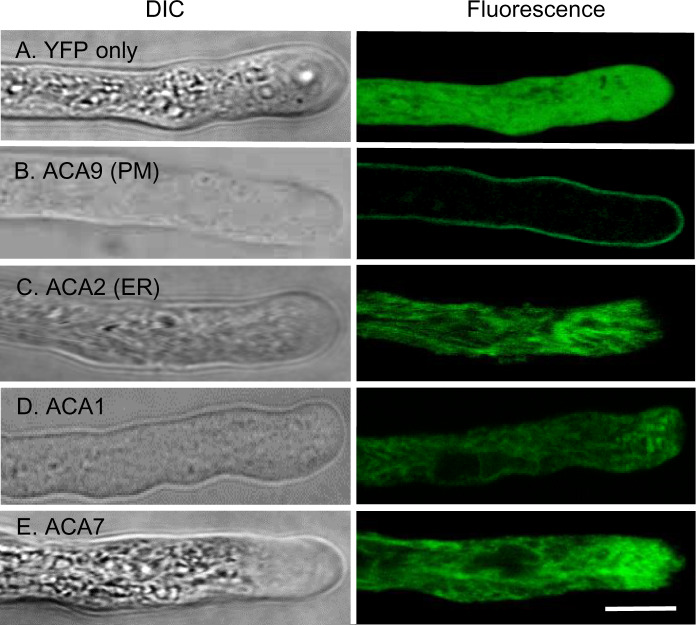
Figure 3.
Confocal fluorescence microscopy of pollen tubes showing FP-tagged ACA1 and 7 with subcellular localizations similar to ER-localized ACA2. Pollen tubes were grown in vitro and imaged using confocal microscopy. Bright field (DIC) and fluorescence are shown. A, YFP shows a positive control for cytosolic localization. B, ACA9–YFP shows a control for PM localization (Schiøtt et al., 2004). C, ACA2-GFP shows a comparison for ER localization (Hong et al., 1999). D and E, ACA1–YFP and ACA7–YFP showing an endomembrane pattern similar to ACA2–GFP. At least two independent lines were analyzed for each ACA–FP fusion. The expression of all fusion constructs were under the control of an ACA9 promoter (Schiøtt et al., 2004). Corresponding ss numbers and ps are YFP only (ss2228 and ss2232, ps532), ACA9-YFP (ss2229–2231, ps580), ACA2-GFP (ss2253–2255, ps585), ACA1–YFP (ss2220–2222, ps1295), and ACA7–YFP (ss2249–2252, ps1960). Scale bar is 10 µm.

