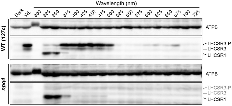
Figure 1.
LHCSR protein levels under different wavelengths of light. Protein was extracted from samples of wild-type (WT; 137c) and npq4-mutant strains. Cells were maintained in darkness (Dark) or treated with 100 μmol photons/m2/s of white light (WL) or different wavelengths of monochromatic light, as indicated, for 4 h. Antibodies against ATPB or LHCSRs (recognizing both LHCSR1 and LHCSR3) were used for immunoblotting analysis. LHCSR3-P represents LHCSR3-phosphorylated. The ATPB protein was used as a loading control. Representative immunoblots from one of three replicated experiments are shown, each performed using different biological samples.

