
Figure 3.
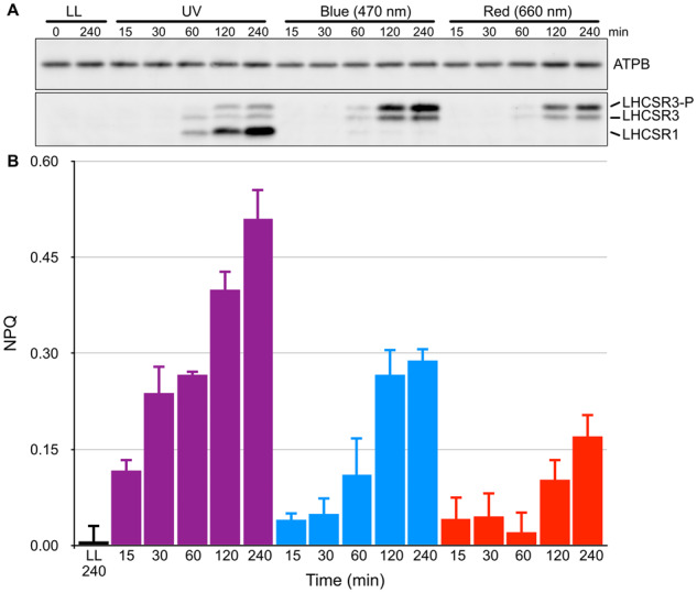
Activation kinetics of LHCSR-dependent photoprotection (NPQ) under different light conditions. A, Wild-type cell samples were collected after 15, 30, 60, 120, and 240 min of irradiation with low-level UV-supplemented fluorescent light at 10 μmol photons/m2/s (Supplemental Figure S3), blue (470-nm) LED light, or red (660-nm) LED light at 110 μmol photons/m2/s, and compared with control samples maintained under LL (30 μmol photons/m2/s) for the duration of the experiment. LHCSR1 and LHCSR3 protein levels were detected using an antibody against LHCSRs (recognizing both LHCSR1 and LHCSR3). LHCSR3-P represents LHCSR3-phosphorylated. The ATPB protein detected using a specific antibody was used as a loading control. Representative immunoblots from one of three replicated experiments are shown, each performed using different biological samples. B, NPQ activities of cells from A measured using a FluorCAM system. NPQ values of cells treated with LL (black bar), UV light (purple bars), blue light (blue bars), and red light (red bars) are shown. Data are mean ± se, n = 3 biological replicates.