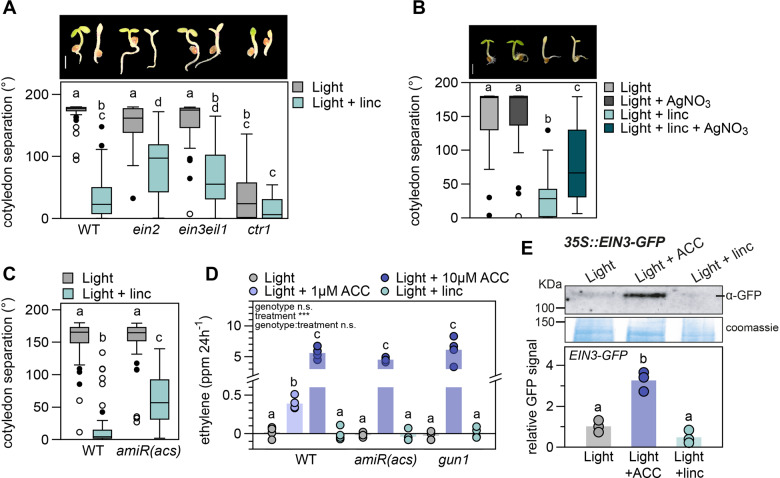
Figure 2.
Lincomycin-mediated repression of cotyledon separation acts via the ET signaling pathway without altering ET emission and ETHYLENE INSENSITIVE3 (EIN3) levels. A, Cotyledon separation (degrees) of 3-d-old, light-grown WT, ein2, ein3eil1, and ctr1 seedlings on regular growth medium or medium supplemented with 0.5 mM lincomycin (linc). Pictures are representative seedlings, scale bar = 1 mm. B, Cotyledon separation of 3-d-old, light-grown WT seedlings on medium supplemented with 5 µM AgNO3, 0.5 mM linc, or a combination of both. Pictures are representative seedlings, scale bar = 1 mm. C, Cotyledon separation of WT and ET-deficient amiR(acs) mutants, grown as in (A). For (A–C), biological replicates n = approx. 40 seedlings. D, ET emission (in ppm/24 h) of 120 WT, amiR(acs) and gun1 seedlings grown in light, on regular growth medium, or supplemented with 1 µM (WT only) or 10 µM ACC, or 0.5 mM linc. Dots represent individual measurements (n = 4), bars are averages. E, EIN3 protein accumulation in 3-d-old 35S::EIN3-GFP/ein3eil1 seedlings grown in low light on regular growth medium, or medium supplemented with 10 µM ACC or 0.5 mM linc, detected by anti-GFP antibodies. Data are relative to light control and Coomassie staining, dots represent biological replicates (n = 3), bars are averages. Complete immunoblot image presented in Supplemental Figure S3A. In (A–E), different letters mark significant differences, p < 0.05 (A and D: two-way ANOVA and post hoc Tukey, B and C: Kruskal–Wallis with post hoc Dunn, E: one-way ANOVA with post hoc Tukey). Images of plants in (A) and (B) were digitally extracted for comparison.

