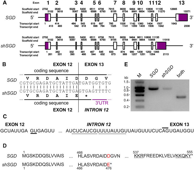
Figure 2.
Alternative splicing generates shSGD by intron retention. A, Genomic organization of SGD and shSGD depicting the general exon/intron junctions. The genomic coordinates were obtained with a reconstructed scaffold from the sequencing of a genomic BAC containing the SGD locus. B, Focus on the 3′-end of exon 12 and on the consequence of intron retention. C, Donor and acceptor sites of splicing of intron 12 are highlighted by solid lines and putative branch points are shown by white rectangles. D, Amino acid sequences of SGD and the truncated protein encoded by SRR924148_TR34256_c6_g1_i5_len=2115. Red letters and stars indicate mutated amino acids and protein ends, respectively. The bipartite NLS of SGD is underlined. E, Amplification of specific fragments of SGD and shSGD transcripts by RT-PCR performed on RNA from leaves. A combination of primers common to both transcripts was used as a control.

