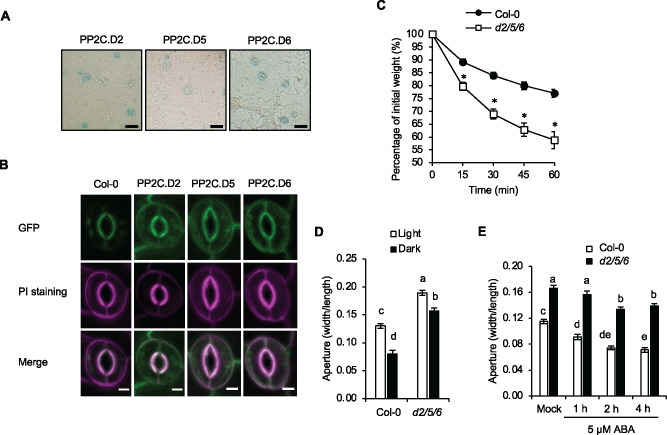
Figure 2.
The pp2c.d2/5/6 triple mutant exhibits increased stomatal aperture diameter. (A) X-Gluc staining of native promoter:PP2C.D-GUS translational fusion proteins in leaf epidermal peels. Scale bar = 25 µm. (B) Subcellular localization of native promoter:PP2C.D-GFP fusion proteins (green) in guard cells of 7-d-old cotyledons. Propidium iodide (PI) (magenta) was used to mark the cell periphery. Scale bar = 5 µm. (C) Water loss in leaf detachment assays of wild-type Col-0 and pp2c.d2/5/6 triple mutants. Data are means ± se (n = 8). Asterisks indicate significant difference from the wild-type (P < 0.05, Student’s t test). (D) Stomatal aperture measurements of wild-type Col-0 and pp2c.d2/5/6 triple mutants during light and dark conditions for 2 h. Data are means ± se (n = 60). (E) Stomatal aperture measurements of the wild-type and pp2c.d2/5/6 triple mutants following treatment with 5 µM ABA. Data are means ± se (n = 60). Different letters above error bars indicate significant difference (P < 0.05) by one-way analysis of variance analysis with Tukey’s HSD test.

