Figure 2.
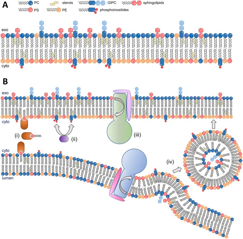
Membrane lipid asymmetry. A, The plasma membrane of eukaryotic cells presents an asymmetric lipid distribution where phosphatidylserine (PS), phosphatidylethanolamine (PE), and phosphatidylinositol phosphates (PIPs) are concentrated in the inner leaflet, and phosphatidylcholine (PC), glycosylinositolphosphorylceramides (GIPCs), and other sphingolipids are concentrated in the outer leaflet. Sterols give the membrane rigidity. B, Several cellular mechanisms coordinate to establish membrane lipid asymmetry. (i) Lipid transfer proteins move lipids between the cytosolic leaflets of two membranes at membrane contact sites. (ii) Phosphatidylinositol kinases and phosphatases phosphorylate/dephosphorylate their targets to generate specific PIPs at the cytosolic leaflet of cellular membranes. (iii) ATP-dependent transporters, such as P4 ATPases (depicted), translocate specific lipids between the two leaflets of the membranes in the secretory pathway. (iv) The activity of P4 ATPases contributes to vesicle budding in the secretory pathway and the subsequent flow of vesicles transports lipids among membranes. Lipids that are synthesized in the lumen of organelles, such as sphingolipids, are confined to the inner leaflet of the lipid bilayer in vesicles and are exposed to the extracellular/lumenal side of the membrane after vesicle fusion to its final destination.

