
Fig. 3. Free radical measurements in single mitochondria.
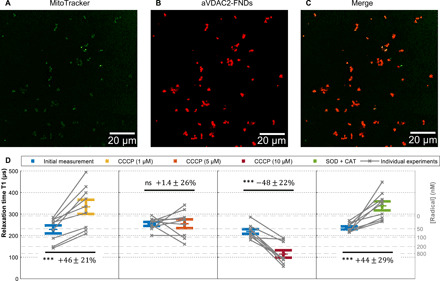
(A to C) Confocal images of aVDAC2-FND targeted to isolated mitochondria. Green, MitoTracker Green–stained mitochondria; red, FNDs. Scale bar, 20 μm. (D) Free radicals detected on single isolated mitochondria by T1 measurements. After recording an initial T1 measurement, 1, 5, and 10 CCCP as well as SOD (1000 U/ml) and CAT (600 U/ml) were tested on the same samples. The error bars represent SEs. The left gray axis represents the approximated concentration determined from a calibration with *OH radicals in solution from the previous work (45). The experiments were repeated nine times. The data were analyzed by using a paired t test against the previous group. ***P ≤ 0.001.