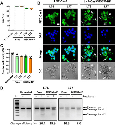
Fig. 3. LNP-Cas9 loaded onto MSCM-NF showed low leukemia cell toxicity, high uptake efficiency, and comparable gene editing efficiency to bolus delivery.

(A) Cell uptake rate of FITC-labeled free LNP-Cas9 or LNP-Cas9 on MSCM-NF after 4 hours of treatment. The FITC-positive population was measured by flow cytometry. Data are means ± SD. P < 0.0001 was observed between free and MSCM-NF groups. One-way analysis of variance (ANOVA) with Tukey’s multiple comparison test. (B) CLSM images showing intracellular uptake of FITC-LNP-Cas9 in leukemia THP-1 cells after 24 hours of treatment. Nucleus was stained with 4′,6-diamidino-2-phenylindole (DAPI). THP-1 cells did not take up the lengthy NF (arrow). Scale bar, 25 μm. (C) Cytotoxicity of free LNP-Cas9 RNP or LNP-Cas9 RNP on MSCM-NF. Cell viability was measured after 24 hours of treatment and normalized to the untreated group. n = 2 independent studies. Data are means ± SD. No significant difference was observed between free and MSCM-NF groups. One-way ANOVA with Tukey’s multiple comparison test. (D) Gene editing efficiency of free LNP-Cas9 RNP and LNP-Cas9 RNP/MSCM-NF in leukemia cells. THP-1 cells were incubated with free LNP-Cas9 RNP or LNP-Cas9 RNP/MSCM-NF for 2 days, and cells were harvested for a gene editing detection assay. Free, Free LNP-Cas9 RNP. MSCM-NF, LNP-Cas9 RNP/MSCM-NF. L, 100-bp DNA ladder.
