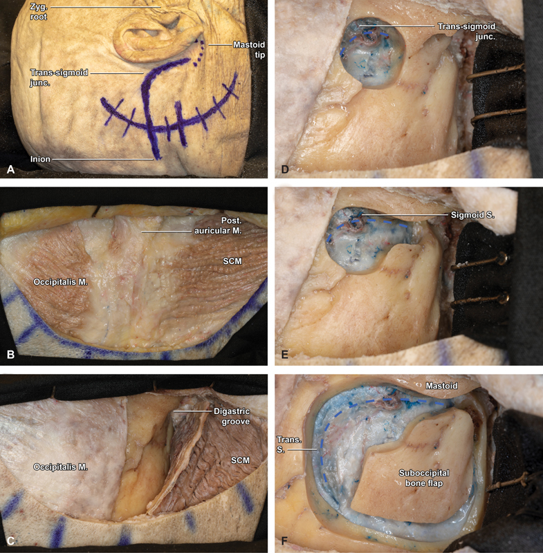
Fig. 1.

Step-by-step retrosigmoid craniotomy in an anatomical specimen (right side). ( A ) Marked skin incision approximately 5-cm posterior to the digastric notch (6-cm posterior to the helix). The transverse sinus is approximated by connecting an imaginary line from the inion to the zygomatic root, while the sigmoid follows the digastric groove. ( B ) With the postauricular scalp flap reflected anterior, the underlying musculature is visualized, including the occipitalis, posterior auricular, and sternocleidomastoid (SCM) muscles. ( C ) Three cuts are made in SCM roughly paralleling the digastric groove, SCM insertion, and medial margin of the skin incision. ( D ) With the SCM flap protected, reflected inferiorly, and secured with 2 fish hooks, a large burr hole is fashioned overlying the transverse sigmoid junction (blue hash). ( E ) Bone removal is carried from the burr hole inferiorly, tracing the course of the sigmoid sinus to the level of the mastoid tip (blue hash). ( F ) After carefully and extensively stripping the dura from the inner table of the occipital bone, a small, rectangular bone flap is turned with the spiral bit and footplate attachment, with the superior and lateral bony exposure revealing the margins of the transverse and sigmoid sinuses (blue hash). Junc, junction; M, muscle; Post, posterior; S, sinus; SCM, sternocleidomastoid; Trans, transverse; Zyg, zygomatic.
