Fig. 2.

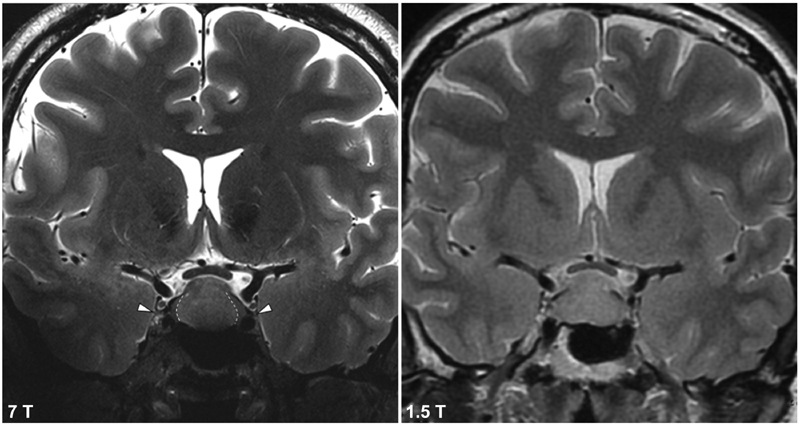
Coronal T2-TSE depicting the trochlear nerve (CN IV) at 7 T (left) and 1.5 T (right). Dashed lines represents demarcation between central tumor or and preserved normal gland laterally. This boundary is not seen at 1.5 T. CN, cranial nerve.

Coronal T2-TSE depicting the trochlear nerve (CN IV) at 7 T (left) and 1.5 T (right). Dashed lines represents demarcation between central tumor or and preserved normal gland laterally. This boundary is not seen at 1.5 T. CN, cranial nerve.