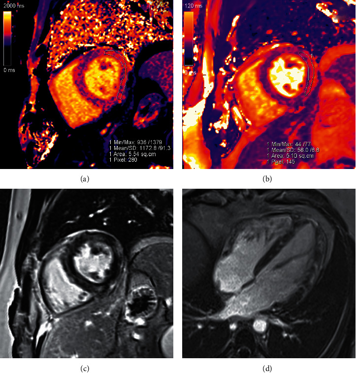
Figure 3.

Cardiac MRI showed features consistent with myocarditis satisfying the updated Lake Louise criteria [4], visualized in these images. (a) Mid-short-axis native T1 mapping demonstrated elevated native myocardial T1 at 1173 ms (local lab normal reference: 950–1050 ms). (b) Mid-short-axis T2 mapping demonstrated elevated myocardial T2 at 58 ms (normal reference: 40–50 ms). (c) Mid-short-axis phase-sensitive inversion recovery late gadolinium enhancement image showed multiple areas of subepicardial enhancement in the lateral wall, as well as midmyocardial enhancement in the septum. (d) Four-chamber late gadolinium enhancement image demonstrated midmyocardial enhancement in the septum and subepicardial to midmyocardial enhancement in the lateral wall.
