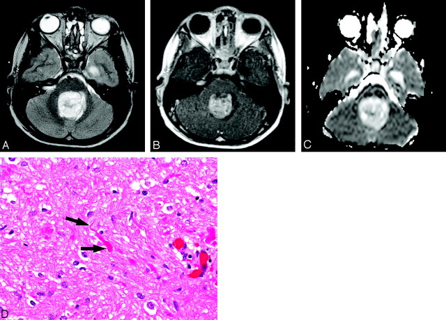
Fig 2.
Eleven-year-old boy with cerebellar juvenile pilocytic astrocytoma (JPA) (patient 4).
A, Axial T2-weighted image at the level of middle cerebellar peduncles shows slightly heterogeneous, predominantly hyperintense midline mass without significant surrounding edema. There is associated effacement of the fourth ventricle.
B, Contrast-enhanced T1-weighted image at same levels as A demonstrates strong, slightly heterogeneous enhancement of the tumor.
C, Apparent diffusion coefficient (ADC) map corresponding to A and B reveals that lesion is very hyperintense compared with normal brain parenchyma, representing increased diffusion of water.
D, Photomicrograph shows area of pilocytic astrocytoma with denser stroma and numerous Rosenthal fibers (arrows) (hematoxylin-eosin stain, 20×).

