Fig 3.
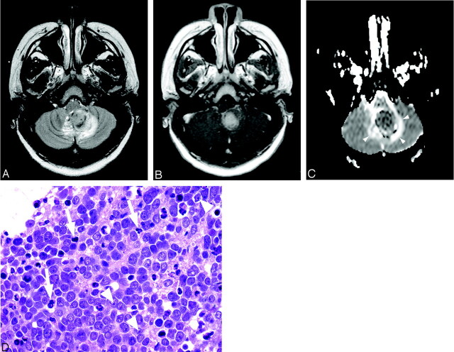
Sixteen-year-old boy with cerebellar medulloblastoma (patient 23).
A, Axial T2-weighted image at level of medulla oblongata shows heterogeneous mass in left paramedian location that is predominantly hyperintense. There is surrounding edema and compression of fourth ventricle.
B, Contrast-enhanced T1-weighted image corresponding to A demonstrates avid, slightly heterogeneous, enhancement of tumor.
C, ADC map corresponding to A and B reveals that mass is hypointense to normal cerebellar parenchyma, consistent with decreased diffusion. Hyperintense ring surrounding tumor (arrowhead) represents increased diffusion of vasogenic edema.
D, Photomicrograph shows densely packed nuclei in medulloblastoma with scattered apoptosis and mitoses (arrows). Some nuclei show prominent nucleoli (arrowheads) (hematoxylin-eosin stain, 40×).

