Fig 7.
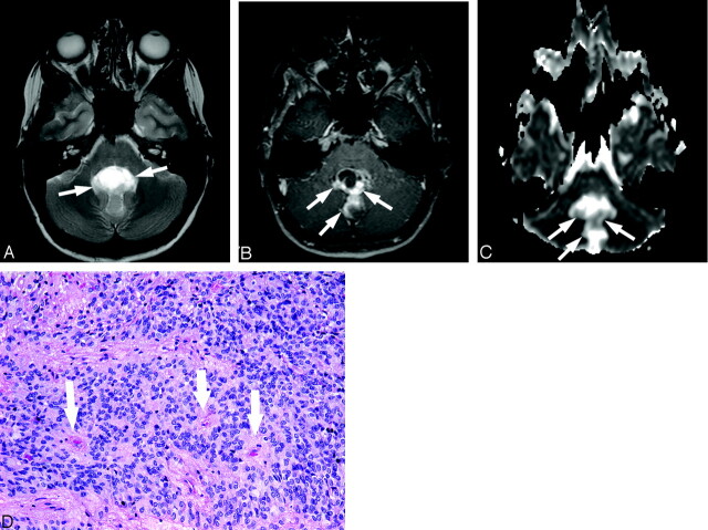
Sixteen-year-old boy with ependymoma (patient 19).
A, Axial T2-weighted image at level of middle cerebellar peduncles shows a very heterogeneous abnormality (arrows) within the fourth ventricle.
B, Corresponding contrast-enhanced T1-weighted image demonstrates enhancement of the solid portion of this mass (arrows).
C, ADC map at a level similar to that of A and B shows that diffusion within the solid portion of the tumor (arrows) is slightly higher compared with normal cerebellum.
D, Photomicrograph of ependymoma shows moderate cellularity with perivascular pseudorosettes (arrows) (hematoxylin-eosin stain, 10×).

