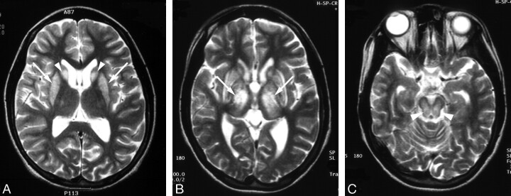
Fig 5.
Wilson disease in a 14-year-old girl with dystonia.
A, Initial T2-weighted axial MR image shows increased signal intensity in both caudate nuclei (arrowheads) and putamen (arrows).
B and C, Follow-up T2-weighted axial MR images obtained after 3 years show aggravated lesions, especially in the thalamus (white arrows) and midbrain (arrowheads). Clinically, the patient’s neurologic symptoms, including dystonia, were deteriorated, despite adequate treatment.

