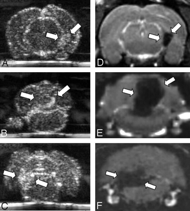
Fig 2.
Comparison of SPIO-enhanced sonographic imaging of EAE lesions with MR imaging. We performed in vivo MR imaging to correlate the sonographic results with a well-established technique to monitor SPIO-particle accumulation. On coronal CISS images (D–F) corresponding to the brain regions depicted by sonography (A–C), we identified areas of focal hypointensity in the periventricular area (A, -D) and in the cerebellar (B, -E) or brain stem region (C, -F) of rat brains (lesions marked by white arrows). Hypointense areas are indicative of local SPIO-particle accumulation. Note the marked similarity of lesion distribution between sonograms and MR images (compare A–C with D–F).

