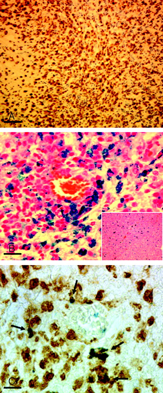
Fig 3.

Histologic evaluation. Paraffin sections were cut through the lesions as detected on MR imaging and sonography. A and B, The histology corresponds to the cerebellar lesion depicted in Figs 1A and 2A. A, Staining of macrophages and activated microglia by immunohistochemistry for ED1 (bar = 50 μm). Note the massive infiltrate of brown-stained ED1–positive cells. On a consecutive section (B, bar = 15 μm), Perls blue staining was performed to detect focal iron accumulation. Note the extensive perivascular iron deposition. Perls staining of liver sections with massively iron-laden Kupffer cells served as a histologically positive control for correct intravenous SPIO-particle injection (B, inset). Combination of Perls staining with ED1 immunohistochemistry reveals a clear colocalisation (C, bar = 10 μm), thereby identifying macrophage-like cells as one of the definitively iron-laden cell types in the CNS.
