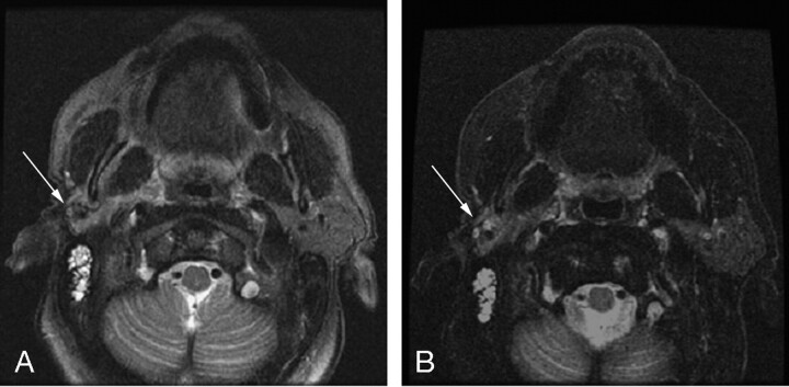
Fig 1.
T2-weighted axial images obtained by using (A) CHESS-FSE sequence with a scan time of 2:32 minutes and (B) FSE-IDEAL sequence with a scan time of 2:33 minutes. The patient had a history of right parotidectomy for poorly differentiated parotid carcinoma. Despite careful shimming, fat signal intensity in the region of the operative bed (arrow) in the CHESS-FSE sequence is not clearly suppressed. The FSE-IDEAL image has uniform fat suppression throughout the field, allowing for better visualization of residual parotid parenchyma (arrow).

