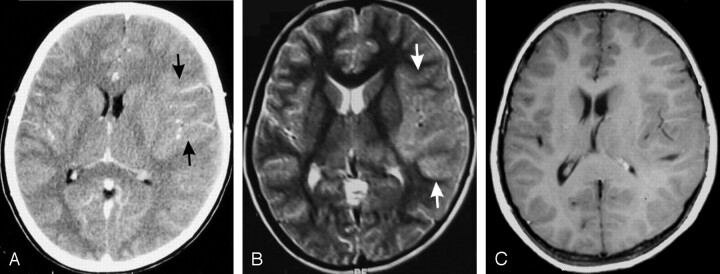
Fig 2.
A 10-year-old boy with headache and vomiting for 2 and a half months (GAE).
A, Contrast-enhanced CT shows an ill-defined pseudotumoral lesion in the left frontotemporal lobe (arrows), with a patchy linear type of enhancement.
B, T2-weighted MR image shows that the mass lesion is isointense to gray matter (arrows). No perilesional edema or any significant mass effect is seen. There were no other focal lesions.
C, Two-month follow-up contrast-enhanced MR image reveals persistence of the mass lesion, with no significant contrast enhancement.

