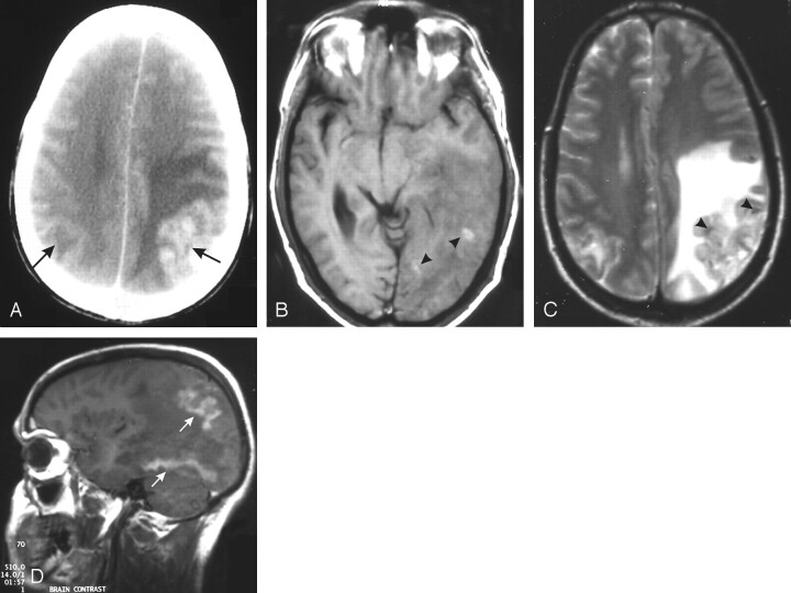
Fig 4.
A 38-year-old man with deteriorating vision and persistent vomiting for a week (GAE).
A, Contrast-enhanced CT scan of the brain shows an enhancing cortical-based lesion in the left parietal lobe with extensive perilesional edema. Another smaller hypoattenuated lesion is seen on the opposite side (arrows).
B, T1-weighted MR image shows hemorrhagic foci (arrowheads) in the left parietooccipital lesion with ill-defined gray-white matter distinction.
C, T2-weighted MR image shows heterogeneous signal intensity in the swollen cortex (arrowheads) with extensive white matter edema. The right-sided lesion also shows mild edema and cortical signal-intensity changes.
D, Contrast-enhanced T1-weighted sagittal image shows gyriform enhancement of the affected cortex with nonenhancing edematous white matter. Similar enhancement is also seen in the undersurface of the occipital lobe (arrows).

