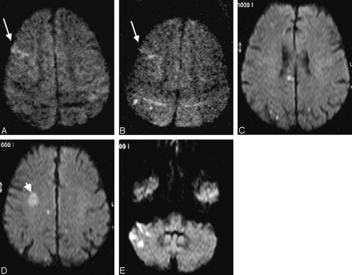
Fig. 2.
Range of expression of diffusion-weighted imaging (DWI) lesions after filter-protected internal carotid artery (ICA) stent placement.
A and B, DWI the day before and the second day after stent placement of a right ICA stenosis. Upper limit of a small cortical infarct in the right middle cerebral artery (MCA) territory is visible on both images (arrow). Typical appearace of 2 new punctate lesions in the right parietal lobe (B).
C–E, Poststent DWI after endovascular treatment of a right ICA stenosis in the patient who showed the maximal number and extension of new lesions in this study. New hyperintensities are detected in the ipsilateral ICA as well as in the contralateral ICA (C and D) and vertebrobasilar territory (E). Note a pre-existing subacute infarct in the right MCA territory (arrowhead, D).

