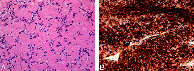
Fig 3.
A, Photomicrograph shows the tumor specimen to be of low cellularity and the presence of cells with oval nuclei arranged in an irregular manner. These are areas consistent with the Antoni A tissue pattern characteristic of schwannoma (hematoxylin and eosin/saffranin, original magnification, ×300).
B, Diffuse reactivity for S-100 protein antibody is present in the specimen. This protein is found in high concentrations within Schwann cells (S-100 stain, ×300).

