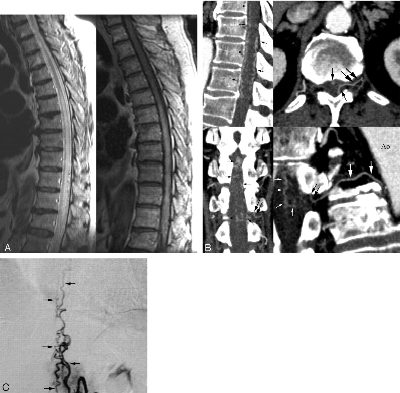
Fig 2.
55-year-old man with SDAVF.
A, Sagittal fast spin-echo T2-weighted MR (left) and postgadolinium T1-weighted images (right) show multiple enlarged pial vessels along the surface of the cord. Intrinsic increased signal intensity centrally within the spinal cord (left) and abnormal enhanced cord (right) extend from midthoracic levels to the conus medullaris.
B, Sagittal (upper left) multiplanar reconstruction image shows multiple engorged perimedullary veins (arrows) along the anterior and posterior surfaces of the spinal cord. Transverse MIP image (upper right) shows the fistula (large arrows) fed by the radiculomeningeal branch of the left T12 intercostal artery and engorged perimedullary veins (small arrows). Oblique coronal multiplanar reconstruction image (lower left) reveals the fistula (large arrows) and extensive engorgement of perimedullary veins (small arrows). Curved planar reformation image (lower right) along the left T12 intercostal artery and SDAVF delineates the aorta (Ao), intercostal artery (large white arrows), fistula (black arrows), and multiple engorged perimedullary veins (small white arrows).
C, Conventional angiography of the left T12 intercostal artery obtained in the arteriovenous phase, anteroposterior view, shows findings similar to those in Fig 2B.

