
Fig 1.
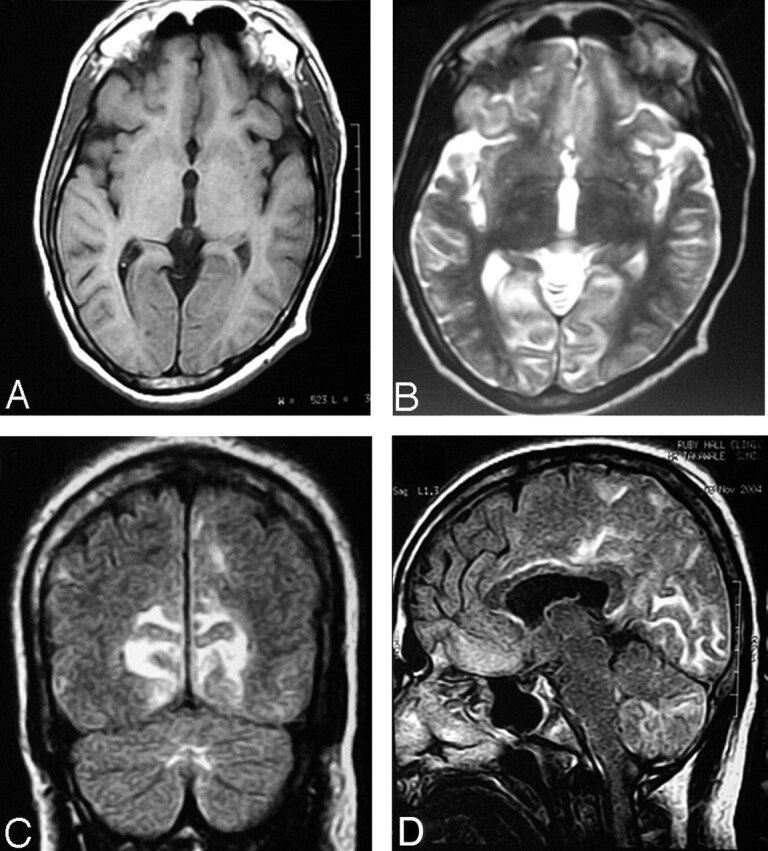
Prechelation MR imaging.
A–C, T1- and T2-weighted axial and FLAIR (respectively) coronal MR images show cortical gray matter and subcortical white matter lesion in the occipital lobe with edema and sulcal effacement.
D, FLAIR sagittal image shows involvement of subcortical white matter in the frontal regions, parieto-occipital lobe, body of the corpus callosum, and cerebellum.