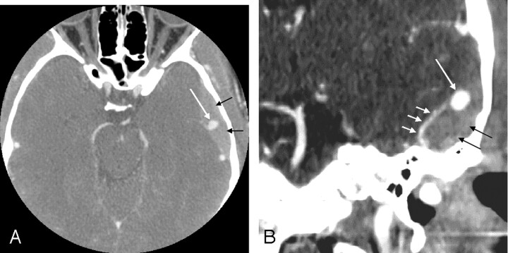
Fig 1.
CT angiogram of the brain in axial plane (A) with coronal maximum intensity projection (MIP) reconstructions (B), done at the initial time of presentation, reveals a pseudoaneurysm (long white arrow) arising from the left middle meningeal artery (MMA) (short white arrows). Left temporal epidural hematoma is also visualized (black arrows).

