
Fig 1.
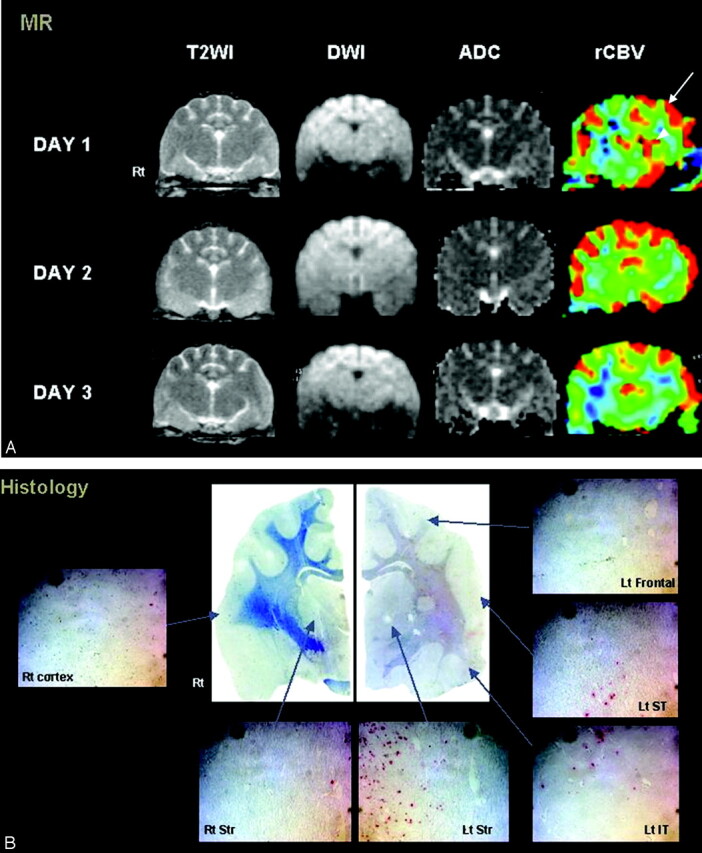
Agmatine-treated cat with type I and II reperfusion.
A, Persistent hyperperfusion is noted in the left frontal lobe (type I reperfusion, arrow). Initial hyperemia in the left striatum shows hypoperfusion on the last follow-up image (type II reperfusion, arrowhead). Notice the lack of signal-intensity changes on T2-weighted (T2WI) or diffusion-weighted images (DWI.). (B) A few red-stained TUNEL-positive cells are seen in all regions of the left hemisphere. ADC indicates apparent diffusion coefficient.