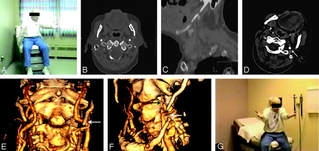
Fig 1.
A, A single captured image of a digital video file shows the patient with head oriented to the left and downward and with the right arm drooping following 25 seconds in this orientation.
B and C, Axial CT image of the neck shows both styloid processes (B); whereas a sagittal reconstruction of the same CT scan shows a calcified stylohyoid complex (C).
D, Axial image from a CT angiogram obtained with the patient’s head in symptomatic orientation demonstrates the calcified styloid process (arrow). No definite flow is seen within the left ICA at this level.
E and F, 3D reconstructions of a CT angiogram obtained with the patient’s head in neutral position (E) show a patent left ICA flowing past the tip of the styloid process (arrow, E), whereas a 3D reconstruction of the CT angiogram obtained with the patient’s head in the symptomatic orientation shows severely diminished flow in the left ICA (black arrow, F) distal to its passage under the stylohyoid complex (white arrow, F).
G, A digitally captured image from a video file obtained after styloid process resection shows no arm droop deficit following 30 seconds in the same orientation as the preoperative picture (A).

