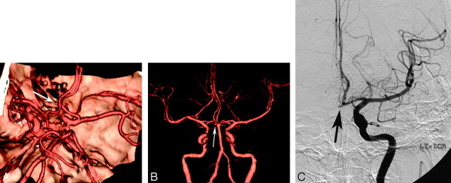
Fig 3.
A 57-year-old man with subarachnoid hemorrhage.
A, Superior projection volume-rendered MDCTA image shows a small aneurysm of the anterior communicating artery (arrow). With this technique, the neck of the aneurysm is obscured by both overlying anterior cerebral arteries despite multiple projections (not shown).
B, Anteroposterior projection volume-rendered MDCTA image with automated segmentation shows laterally and inferiorly directed saccular aneurysm at the anterior communicating artery (arrow). The relations of the aneurysmal neck can be demonstrated better with this postprocessing software.
C, Corresponding DSA image of the left internal carotid artery shows the same aneurysm (arrow).

