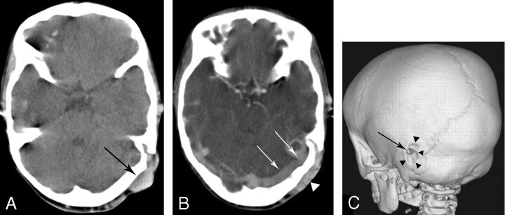
Fig 1.
CT scan of the patient.
A, Noncontrast axial CT scan shows subcutaneous mass (arrow) and local skull defect on the upper portion of the petrous bone.
B, Contrast axial CT scan shows that the enhanced subcutaneous mass (arrow) is communicated with the lateral sinus (arrowheads) through the skull defect, which suggests that the mass lesion is dilated scalp veins filled through the prominent mastoid emissary foramen.
C, 3D CT-shaded surface display showing enlargement of the outer opening of the mastoid emissary foramen (arrow) and depression of the skull surface (arrowheads) around it.

