
Fig 1.
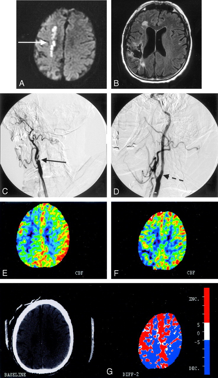
A, Diffusion-weighted MR imaging of patient 1 shows a linear pattern of increased signal intensity in the internal border zone region of the right hemisphere (white solid arrow). B, Fluid-attenuated inversion recovery image sequence shows an old right parietal infarct with encephalomalacia that was clinically silent before the procedure. C, Conventional angiography from the right common carotid artery confirms the presence of a total occlusion of the right internal carotid artery at the bifurcation (solid black arrow). D, Poststent placement and angioplasty of the occlusion show normal antegrade flow distal to the previously occluded segment (dashed black arrow). E, A xenon CT postacetazolamide performed before stent placement shows lack of cerebral vasoreactivity in the right hemisphere in contrast to the left side. F, Poststenting and angioplasty xenon CT reveal augmentation of flow to the right hemisphere except for the known area of infarct in the right parietal lobe. G, Color maps demonstrating the differences in flows before (from Fig E) and after (from Fig F) the procedure show the improved augmented blood flow to the right hemisphere as demarcated by the red color.