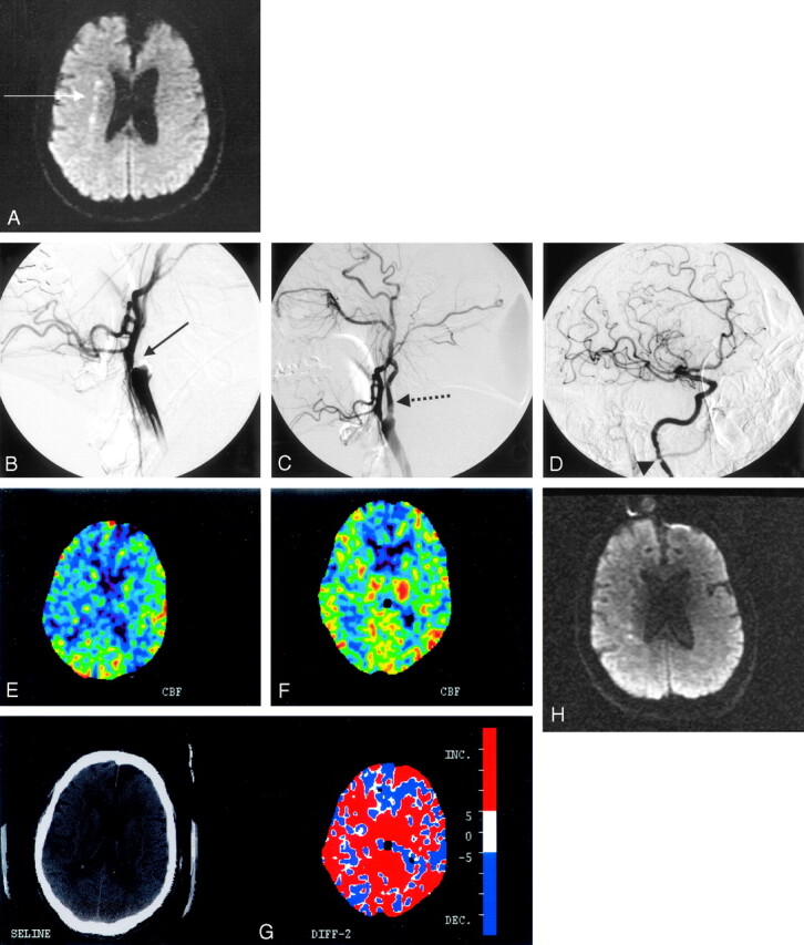
Fig 2.

A, Diffusion-weighted MR imaging of patient 2 shows a linear pattern of increased signal intensity in the internal borderzone region of the right hemisphere (white arrow). B, Conventional angiography confirms the presence of a heavily calcified bulb with total occlusion of the internal carotid artery at the bifurcation (solid black arrow). C, Poststent placement and angioplasty show the antegrade flow across the occluded segment distally (dashed black arrow). D, The patient also had an atherosclerotic stenosis evident at the C1 level of the extracranial internal carotid artery that was not revascularized (black arrowhead). There is normal filling of the intracranial vessels. E, A xenon CT after preprocedure acetazolamide administration demonstrates poor cerebral vasoreactivity to the right hemisphere. F, A xenon CT after postprocedure acetazolamide administration demonstrates robust augmentation of blood flow to the right hemisphere. G, The color map demonstrates the difference in blood flows between Figures E and F, with red demarcating an increase in flow postprocedure. H, Diffusion-weighted MR imaging after stent placement revealed no additional acute infarcts as a result of the procedure.
