
Fig 5.
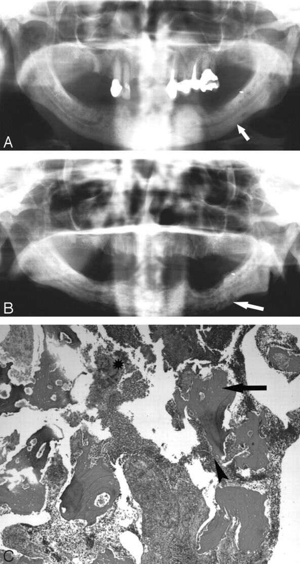
A 77-year-old woman (patient 4) with multiple myeloma initially treated with pamidronate and subsequently zoledronate.
A, The initial orthopantogram demonstrates osseous sclerosis (arrow).
B, Orthopantomogram 9 months later demonstrates lytic destruction of the mandible, most prominent in the left body of the mandible (arrow). Surgical debridement was performed.
C, Photomicrograph at low power (×40) of the curettage specimen stained with hematoxylin-eosin demonstrates fragments of necrotic bone with empty lacunae (arrow). There is extensive infiltration with inflammatory cells with surface resorption of bone (arrowhead) and bacteria (*).