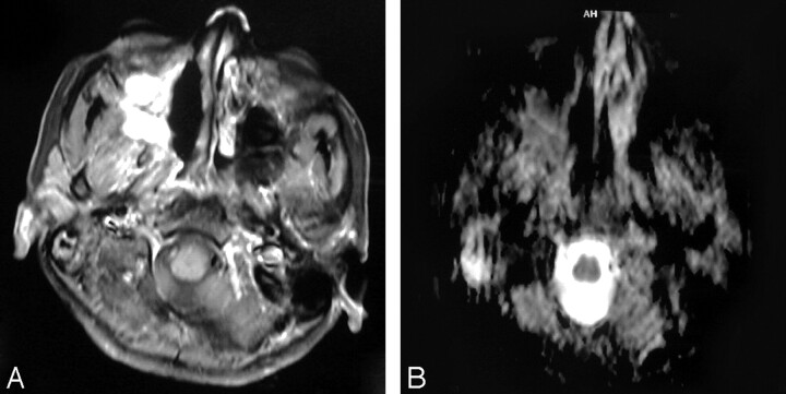
Fig 3.
Recurrent squamous cell carcinoma of the nasal cavity.
A, Axial postcontrast T1-weighted MR image shows that enhancing lesion is seen in the right side of the nasal cavity. Recurrent tumor cannot be differentiated from postradiation changes.
B, ADC map shows hypointensity within the lesion with a low ADC value (1.17 ± 0.17 × 10⁻³ mm2/s).

