Fig 4.
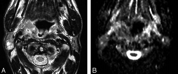
Recurrent squamous cell carcinoma of the oropharynx with metastatic cervical lymph node.
A, Axial T2WI shows an ill-defined irregular mass of inhomogeneous signal intensity involving the right side of the oropharynx. An enlarged cervical lymph node (arrow) with inhomogeneous high signal intensity is also noted at the right side of the neck.
B, ADC map shows low signal intensity of both the lesion and the lymph node with a mean ADC value of 1.20 ± 0.22 × 10⁻³ mm2/s and 1.05 ± 0.20 × 10⁻³ mm2/s, respectively, suggestive of tumor recurrence with metastatic lymph nodes. This was proved by biopsy.

