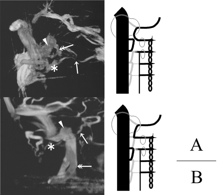
Fig 11.
3DRVs (A, superomedial view of the left side; B, medial view of the left side) and schemas presenting our theory of the development of the caudal end of the IPS (ventral view of the right side) are shown. In these cases, we can find a duplicated venous channel (double arrow) with the IJV. The arrow indicates the IPS, the arrowhead indicates the JB, and the asterisk indicates the ACV.
A, The duplicated channel may be formed with the longitudinal anastomosis of the hypoglossal vein and the 1st cervical intersegmental vein.
B, The duplicated channel may be formed with the longitudinal anastomosis of the 1st and 2nd cervical intersegmental veins.

