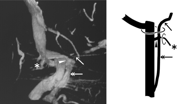
Fig 5.
3DRV (anterolateral view of the right side) and schema (frontal view of the right side) of type C-2: The IPS (arrow) shows a long extracranial extension (double arrow). Note another upper junction (arrowhead) to the IJV and the anastomosis with the VVP via the ACV (asterisk) at the level of the extracranial opening of the hypoglossal canal.

