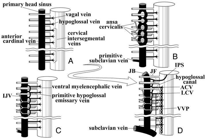
Fig 9.
Theoretic schematic representation of the development of the caudal portion of the IPS in the embryonic and fetal periods.
A, In the early embryonic phases, the primary head sinus and the anterior cardinal vein (the future IJV) run ventromedially to the vagal, accessory, and hypoglossal nerve and to cervical nerve roots.
B, The primary head sinus and the anterior cardinal vein migrate dorsolaterally to the lower cranial nerves and the ansa cervicalis, forming a venous plexus around nerves.
C, The vagal vein, the hypoglossal vein, and the upper cervical intersegmental veins are stretched and anastomose with each other on the ventromedial side of the IJV. The ventral myeloencephalic vein and the primitive hypoglossal emissary vein are formed through this process.
D, The ventral myeloencephalic vein is connected to the cavernous sinus with its cranial end, and the IPS is formed. The morphologic variation of the caudal portion of the IPS may be determined by the extent of development and degeneration of individual venous channels.

