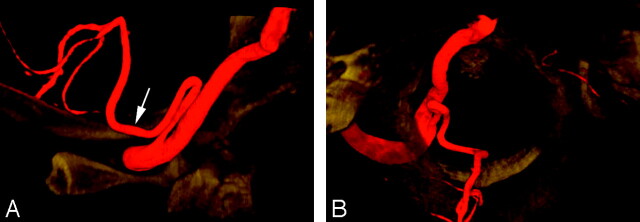
Fig 3.
A proximal (C1) origin of the left PICA (PSA type) in a 47-year-old woman, as demonstrated by a 3D reconstruction of a rotational angiogram with simultaneous osseous and vascular rendering. This technique is known as 3D fusion DSA (3D-FDSA) (Infinix NB, Toshiba, Japan).
A, 3D-FDSA, left VA, lateral view, showing that the proximal portion of the PICA follows the typical course of a PSA trunk as described by Maillot and Koritke7 (ie, a first segment closely paralleling the distal VA, followed by a sharp caudal and dorsal curve that brings the vessel in a posterior-lateral position within the foramen magnum). The ascending ramus of the PSA (white arrow) then continues as the proximal portion of the PICA.
B, 3D-FDSA, left VA, superior axial view, demonstrating the posterior location of the PICA within the foramen magnum. The topographic correspondence between the adult distal VA and PSA/PICA, and the embryonic ventral and dorsal radicular branches of C1 is well illustrated.

