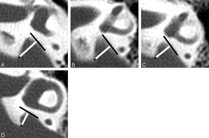
Fig 4.
Technique of measuring the width of the VA at the operculum. Asterisks (*) mark the opercular margins of the VAs. The widths (white lines) are measured from the opercular margins to the spots on the posterior temporal bone walls whose surface is perpendicular to the measurement lines. Tangents (black lines) to these spots are shown to illustrate these right-angle relationships. The magnification of all images is the same.
A, VA opercular width is 5.3 mm.
B, VA opercular width is 4.0 mm.
C, VA opercular width is 4.0 mm.
D, VA opercular width is 3.7 mm.

