Fig 3.
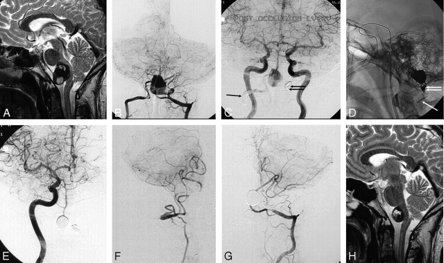
A 29-year-old man with sudden neck pain followed by right-sided muscle weakness and difficulty in swallowing. A and B, MR image (A) and frontal bilateral vertebral angiogram (B) show a giant partially thrombosed vertebrobasilar junction aneurysm compressing the brain stem. C, Bilateral frontal carotid angiogram after occluding the right vertebral artery proximal to the PICA with a balloon (arrow) and the left vertebral artery distal to the PICA with coils (double arrow). Flow to the basilar artery is reversed with outflow to the right PICA, yet the aneurysm lumen still fills. D, Lateral radiograph during coiling of the aneurysm lumen via the posterior communicating artery 2 months later. The arrow indicates deflated balloon remnant in the right vertebral artery. The double arrow indicates coils in the left vertebral artery. E–G, Six months later, a frontal view of right carotid angiogram (E) demonstrates filling of the basilar artery via the right posterior communicating artery. Frontal view of the right thyrocervical trunk (F) shows recanalization of the distal right vertebral artery with filling of the PICA territory. Frontal view of the left vertebral angiogram (G) shows filling of the left PICA territory. The aneurysm is completely occluded. H, MR imaging 2 years after presentation shows remarkable shrinkage of the aneurysm. The patient was free of symptoms.

