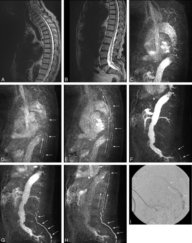
Fig 1.

A and B, Sagittal T2-weighted sequence in case 4 demonstrates serpiginous flow voids along the ventral and dorsal spinal cord from the cervical region to the sacrum. Increased T2-signal intensity is seen within the distal cord/conus. These findings are highly suspicious for a fistula, the level of which cannot be determined on these conventional images. The patient has incidental low-lying conus. C–E, Three MIP partitions from a sagittal TRSMRA (temporal resolution = 5.07 seconds) centered in the cervical and upper thoracic spine show early filling of venous structures on the ventral and dorsal surface of the cord (short arrows). Filling proceeds from caudal to cephalad (long arrows). F–H, Three MIP partitions from a sagittal TRSMRA (temporal resolution = 5.07 seconds) centered in the thoracic, lumbar, and upper sacral spine demonstrate early venous filling (short arrows) beginning within the pelvis and proceeding cephalad (long arrows). Unsubtracted images (not shown) showed the earliest venous drainage within the sacrum. A coronal oblique acquisition further localized the fistula to approximately the S3 level. I, Lateral projection from a superselective catheter spinal DSA shows early venous filling within the sacral canal proceeding cephalad compatible with a spinal DAVF. This correlates with the appearance and physiologic dynamics demonstrated in F–H.
