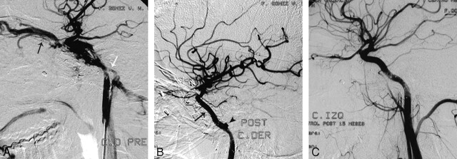
Fig 1.
A, Patient 2. Selective left ICA angiogram demonstrates a CCF after a gunshot wound, with filling of the superior ophthalmic vein (black arrow) and inferior petrosal sinus (white arrow). B, Immediate control post-covered stent deployment (black arrow) shows complete occlusion of the fistula. A small pseudoaneurysm is noticed in the petrous carotid artery (black arrowhead), which was managed conservatively. C, Follow-up after 15 months shows a normal artery without recanalization of the fistula. There has been spontaneous resolution of the small pseudoaneurysm, and there is no intimal hyperplasia.

