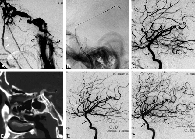
Fig 3.
A, Patient 5. Selective right ICA angiogram demonstrates a high-flow CCF with arterial phase enhancement of a markedly enlarged superior ophthalmic vein (black arrow), facial veins (white arrowheads), and both inferior petrosal sinuses (curved arrows). This is a case of near-complete cavernous ICA transection with nonvisualization of the right anterior and middle cerebral arteries due to complete deviation of the flow into the fistula. B, There is an exchange wire stabilized in one of the distal middle cerebral artery branches. A bare stent has just been deployed in the cavernous carotid artery to reconstruct the vessel wall and provide stability to the covered stent, which is being positioned inside the bare stent at the exact location of the fistula. C, Control postdeployment shows occlusion of the fistula with re-establishment of the intracranial flow through the right ICA. There is straightening (arrow) of the cavernous ICA with no hemodynamic consequence in the control angiogram. D, Three-month follow-up with CT angiography. Here the sagittal reformat shows the straightening of the cavernous carotid artery with the stent in place and preserved patency. E and F, Angiographic follow-up at 8 and 15 months demonstrates a normal intracranial ICA without recurrence of the fistula or significant intrastent intimal hyperplasia (black arrows).

