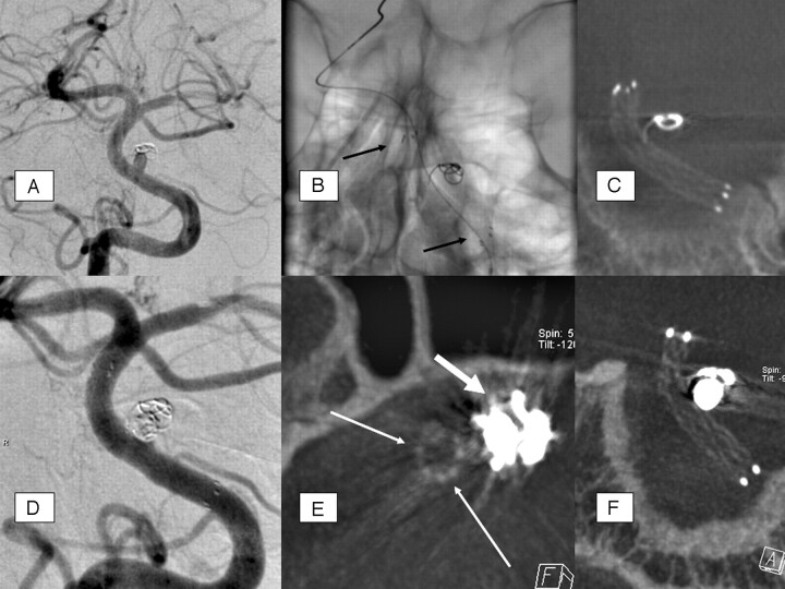
Fig 1.
A, The basilar stem aneurysm showed complete occlusion initially after insertion of a single coil 2.5 mm in diameter: coil compaction and significant aneurysm growth in 6-month follow-up-DSA. B, Unsubtracted DSA image after insertion of a Neuroform stent (4 × 20 mm) shows the proximal and distal radiopaque markers of the stent (arrows); the stent itself is not visible. C, MIP reconstruction of ACT performed after stent deployment and before second coil embolization: excellent stent visibility and regular, complete stent deployment. The initially inserted single coil shows compaction and is distant to the parent (basilar) artery and to the stent struts as well, including one coil loop with position adjacent to stent wall, with definite extraluminal position. D, DSA. A total of 4 platinum microcoils (diameters, 2.5 and 2.0 mm, respectively) were additionally inserted. The basilar stem aneurysm shows satisfiable occlusion with minimal dog-ear remnant. E, Axial, thin (1 mm section thickness) MPR reconstruction of ACT performed after stent deployment and second coil embolization (same dataset as in F). This reconstruction allows definite exclusion of stent strut movement or coil protrusion: stent (thin arrows) without deformation adjacent to coil package (thick arrow), which causes some beam-hardening artifacts. F, MIP reconstruction (oblique coronal) of ACT performed after stent deployment and second coil embolization: excellent stent visibility without significant limitation by marginal beam-hardening artifacts through coil package; no change of stent configuration compared with the ACT imaging before the second coil embolization.

