Fig. 3.
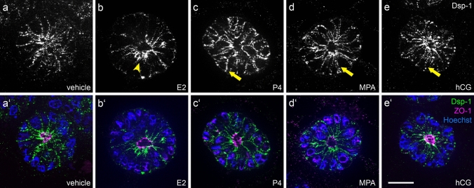
Influence of ovarian steroid hormones and hCG on localization of desmoplakin 1. Images show Ishikawa spheroids after 4 days of stimulation with estradiol/E2 (b/b’), progesterone/P4 (c/c’), medroxyprogesterone acetate/MPA (d/d’) or human choriogonadotropin/hCG (e/e’). a/a’ show the control without hormones. Arrowhead highlights subapical accumulation of Dsp-1 expression (b). Arrows highlight Dsp-1 redistribution to the basolateral membrane (c and d). Grey-scale pictures for Dsp-1 (a–e) or in green combined with tight junctional staining (ZO-1, magenta) and Hoechst (blue) in (a’–e’). Scale bar: 20 µm

