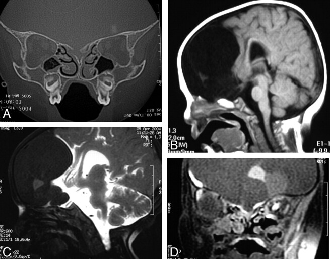
Fig 2.
Dysraphic state with hypertelorism, intracranial suspected dermoid tumor, and bifid nose (patient 8). Coronal CT scan through the cribriform plate (A) and sagittal SE T1-weighted MR image through the anterior cranial fossa (B) do not exclude associated encephalocele. Intrathecal Gd-DTPA-enhanced T1-weighted SE fat-saturated images in sagittal (C) and coronal (D) planes show integrity of the anterior cranial fossa and absence of meningoencephalic herniation. Surgery was delayed without the fear of unnoticed sac rupture.

